
Visualization of internal gas spaces with synchrotron X-ray micro-tomography
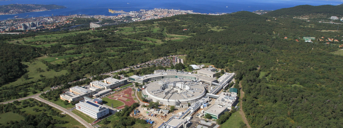
ELETTRA with the town of Trieste in the background. (c) Lightsources.org
The objective of our visit to ELETTRA was to use synchrotron X-ray phase contrast micro tomography (PHC microCT) to visualize tissue bottlenecks for gas-phase diffusion in two wetland plant species showing contrasting tissue O2 dynamics. The visualization of internal networks of gas spaces will resolve the putative tissue bottlenecks in two species of aquatic plants with high tissue porosities (gas spaces) in most of the leaf, stem and root tissues. The visualization will be used to model internal O2 diffusion from shoot to roots, and model calibration is aided by empirical O2 dynamics obtained by O2 microsensors.
The two model plants used were Limonium narbonense and Sarcocornia fruticosa since we have already published a paper where these species show contrasting responses in internal aeration to partial and complete submergence. See more in:Pellegrini E, Konnerup D, Winkel A, Casolo V, Pedersen O (2017) Internal tissue aeration in two halophytes of the Mediterranean region, during partial and complete submergence. Functional Plant Biology 44: 867-876. 10.1071/FP16369