

New paper on methods to visualize or quantify radial O2 loss from plant roots
This paper is a joint contribution from the Nagoya University, Udine University and the University of Copenhagen summarizing methods for visualization and/or quantification of radial oxygen loss (ROL) from plant roots.
We are highlighting four methods, which we think are of key importance: i) the visualization of ROL using methylene blue staining, ii) quantification using the root-sleeving electrode, iii) quantification using an oxygen microsensor and iv) the planar optode to visualize and quantify ROL in 2D.
We hope that you will find the protocols particularly useful; for four of the key methods, these are made available as supporting information. Please do not hesitate to contact us if you have any questions when setting up measuring systems in your own lab.
The paper is OPEN ACCESS and can be accessed from here.
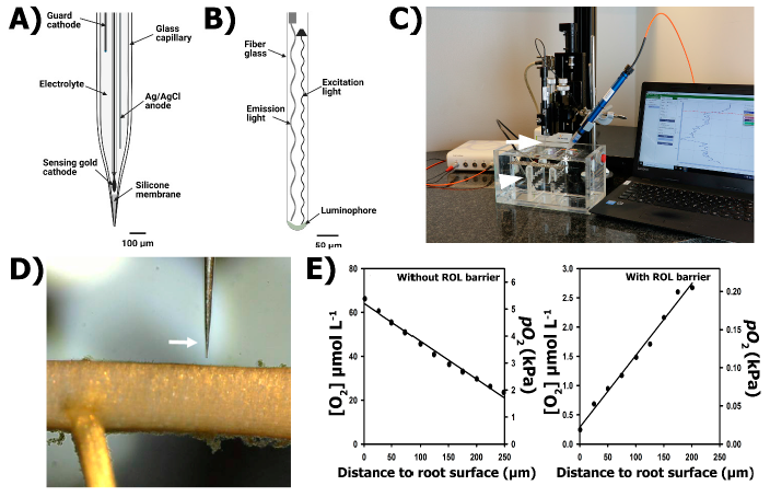
Here is an example of a figure from the paper describing the microsensor technique. For this approach, you need a minaturized Clark-type oxygen sensor or an oxygen optode. In both case, the tip diameter should be 50 µm or less in order to obtain a reasonable spatial resolution of the concentration profile, which is subsequently used to calculate the oxygen flux out of the root, or in some special cases into the root.